one History of canine diseases
The patient is a male Teddy dog, 8 years old and weighing 7kg. When the pet owner brought the pet to the hospital, he reported that he found subcutaneous bleeding and blood spots on the lower abdomen when the pet was bathed at the pet store . The pet owner believed that the pet store used too much force to cause the bleeding. The pet store staff brought the pet owner and the pet to the veterinary hospital for treatment.
The dog owner reported that the dog's defecation and urination had been normal recently. The day before, when the dog was brushed with animal toothpaste, it swallowed a large amount of toothpaste. The dog's feces were black the day before.
Routine examination of the affected dog: body temperature 40.4℃, respiratory rate 50 breaths/min, heart rate 100 beats/min, MMC blue-purple, CRT 1-2s, BCS score 4/5, pet's mood stable and calm.
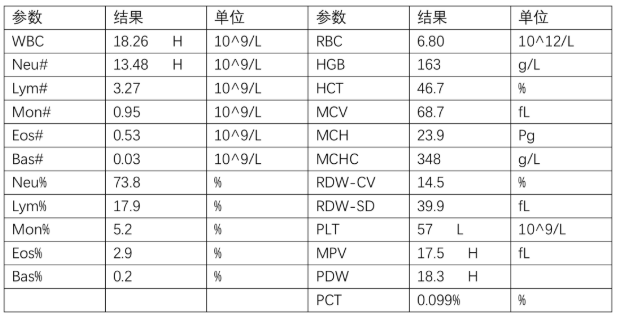
two Complete blood count (CBC) five-part differential test
The disease process disrupts the balance of platelet production, activation, and clearance, leading to an increase or decrease in platelet count. Thrombocytopenia is one of the most common acquired hemorrhagic diseases in small animal clinics, while thrombocytosis is more often considered an abnormal finding on laboratory tests without specific clinical symptoms.
three Crp: CRP level was 16 mg/L, which is elevated.
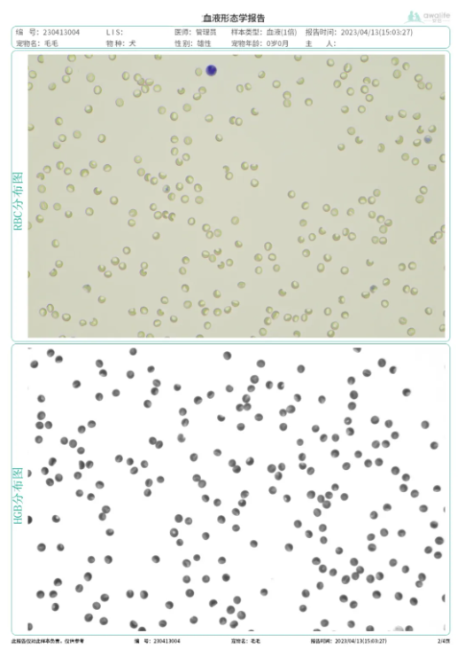
Four Anlu Formed Components Analyzer for Blood Morphology
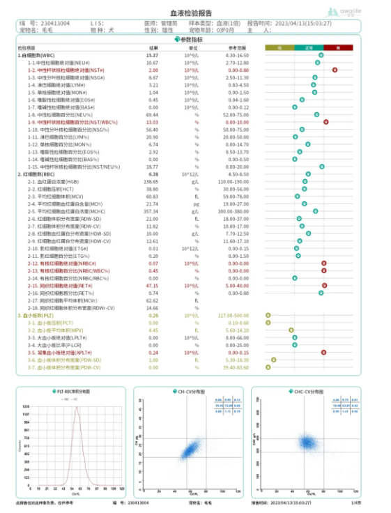
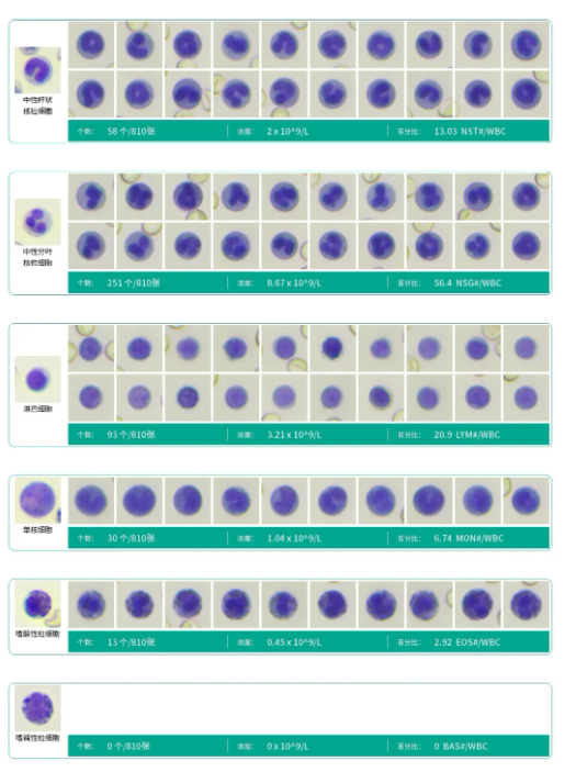
The report shows
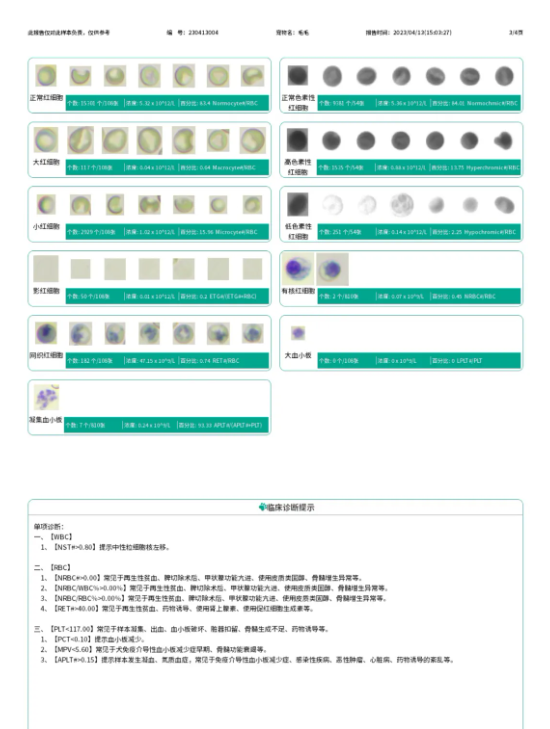
An increase in both the absolute number and proportion of band neutrophils beyond the normal range suggests a left shift of the nuclei; while the total number of neutrophils did not increase significantly, suggesting possible early acute inflammation .
The absolute number of nucleated erythrocytes and reticulocytes increases, indicating a strong regeneration response.
A severely reduced plateletcrit (PCT) and mean platelet volume (MPV) (close to zero) suggest possible platelet consumption and bleeding .
Based on the above blood morphology analysis, the sample shows early acute infection, platelet depletion combined with a regenerative response, suggesting a possible recent bleeding episode. Combined with the owner's mention of a history of melena and the absence of other signs of trauma or bleeding during the examination, gastrointestinal bleeding is suspected.
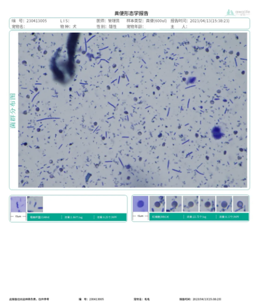
five Anlu Formed Components Analyzer for Stool Examination
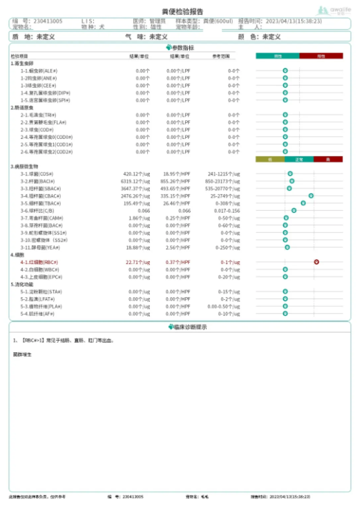
Fecal formed element analysis report diagnosis
After ruling out parasitic and intestinal bacterial infections, the elevated red blood cell count suggests gastrointestinal bleeding . Image verification confirms the presence of a large number of red blood cells.
six Combined diagnosis
The examination included routine blood tests, blood component analysis, fecal component analysis, CRP, combined with the dog's medical history, lifestyle history, recent medication history, and clinical examination.
1. Gastrointestinal bleeding in the affected dog: Combined with the dog's history of black stool, decreased platelet count, increased platelet aggregation, and the discovery of a large number of red blood cells in the fecal examination and morphological examination using a fecal analyzer, it indicates that the dog has gastrointestinal bleeding.
2. The dog is in the early stage of acute inflammation: Based on the CRP results, the total number of neutrophils is not significantly increased, but the number of band neutrophils is increased, indicating that the bleeding has led to bacterial infection in the digestive tract, and the dog is in the early stage of acute inflammation.
seven discuss
In this case, initial CRP and five-part differential blood tests indicated bacterial infection and inflammation in the dog. Red blood cell counts were normal, with no anemia present . The five-part differential blood test showed a slight decrease in platelets, which was not significant and could be due to platelet aggregation.
Blood samples were analyzed using an Anlu formed element analyzer, including six-part differential, reticulocyte, and morphological examinations. The total white blood cell count was not significant, but the total number and proportion of band neutrophils were significantly increased, indicating the dog was in the early stages of inflammation. Red blood cell and hemoglobin levels were within standard ranges, but the reticulocyte count was significantly increased. Reticulocytes are an important indicator of aplastic anemia, suggesting that the dog's blood loss was recent and that the replenishment and release of reticulocytes could meet the body's current oxygen requirements.
In the analysis of formed elements, platelets were almost zero, and there was severe consumption of surface platelets. Bleeding factors were the primary consideration. Morphological examination and image verification confirmed the absence of platelets.
Analysis of the formed elements in the feces revealed a large number of red blood cells, confirming that the dog was currently experiencing gastrointestinal bleeding.
In this case, the blood and stool analysis by the Anlu Formed Component Analyzer enabled the early detection of inflammation progression and bleeding. Furthermore, the combined blood and stool analysis corroborated each other in the diagnosis of gastrointestinal bleeding and infection, thus improving the accuracy of the diagnosis.